Revolutionary 3D Color Imaging Lets Doctors See Inside Your Body Like Never Before
Scientists have developed breakthrough imaging technology that combines ultrasound and photoacoustics to create stunning 3D color visualizations of internal anatomy and blood flow. This could transform how doctors diagnose diseases by providing unprecedented detail of tissue structure and blood-vessel function.
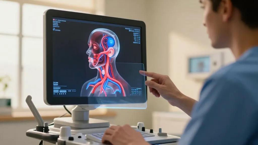
Medical imaging just got a major upgrade that could revolutionize how doctors peer inside the human body. Scientists have developed a groundbreaking technique that combines two powerful imaging methods to create stunning 3D color visualizations of internal anatomy—giving physicians an unprecedented view of both tissue structure and blood flow in real-time.
This innovative approach merges ultrasound technology with photoacoustic imaging, creating a hybrid system that captures details previously impossible to see with traditional medical scans. The result is a vivid, three-dimensional color map that reveals not just what’s inside your body, but how blood moves through your vessels and tissues.
How This Revolutionary Technology Works
The breakthrough lies in the clever combination of two established imaging techniques. Ultrasound, which doctors have used for decades, excels at showing tissue structure by bouncing sound waves off internal organs. Photoacoustic imaging, a newer technology, uses light pulses to visualize blood vessels and monitor blood flow.
By blending these two methods, researchers have created something entirely new—a imaging system that captures both structural anatomy and functional blood vessel activity simultaneously. The technology produces detailed 3D color images that show not just where tissues and organs are located, but how blood circulates through them in real-time.
What Makes This Different from Current Scans
Traditional medical imaging often requires doctors to piece together information from multiple scans to get a complete picture. MRI shows soft tissues well but struggles with blood flow. CT scans reveal bone and some soft tissues but require radiation exposure. Ultrasounds are safe and real-time but typically show limited detail in black and white.
This new hybrid approach addresses many of these limitations by providing:
- Real-time 3D visualization in full color
- Simultaneous tissue structure and blood flow imaging
- Non-invasive scanning without radiation exposure
- Enhanced detail compared to traditional ultrasound
Potential Game-Changer for Medical Diagnosis
The implications for patient care could be significant. Doctors may soon be able to spot diseases earlier and monitor treatment progress more effectively. The technology’s ability to visualize blood vessel function could be particularly valuable for diagnosing cardiovascular conditions, cancer, and other diseases where blood flow patterns provide crucial diagnostic clues.
Reports suggest this enhanced visualization could help physicians identify problems that might be missed with current imaging methods. The real-time nature of the technology also means doctors could potentially guide procedures more precisely while watching both tissue structure and blood flow simultaneously.
What to Watch For
As this technology develops, several key factors will determine its impact:
- Clinical trial results and safety validation
- Cost compared to existing imaging equipment
- Training requirements for medical staff
- Integration with current hospital systems
- Regulatory approval timelines
The Road Ahead
While the technology shows tremendous promise, observers note that significant development work likely remains before it reaches hospital rooms. The research represents an important proof of concept, but clinical trials and regulatory approval processes will determine when patients might benefit from these enhanced imaging capabilities.
The medical imaging field has seen steady advances over recent decades, from the introduction of MRI to the development of 3D ultrasound. This latest breakthrough continues that trajectory, potentially offering doctors and patients a clearer window into human anatomy than ever before.
As healthcare continues embracing technological innovation, imaging advances like this could reshape how diseases are diagnosed and treated, ultimately leading to better outcomes for patients worldwide.